
ALZHEIMER'S DISEASE · SLEEP · EARLY DETECTION

More than 500 healthy people were recorded overnight. The ones carrying the highest genetic risk for Alzheimer's were waking up more often, briefly, without ever knowing it.
Sleep disturbances are increasingly recognized as early features of Alzheimer’s disease (AD) neuropathology. Specifically, spontaneous arousals during sleep have been associated with the burden of Amyloid beta in the brain of healthy late middle-aged individuals. However, it remains unclear whether heterogeneity of arousals relates to genetic risk for AD in younger adults or to cognitive change later in life. Here, we evaluated the association between arousals, polygenic risk scores (PRS) for AD, and cognitive performance and change in healthy young and late-middle-aged individuals.
A team in Liège tracked sleep in over 500 healthy adults using brain recordings, counting micro-awakenings. These are brief flickers of arousal that fragment the night without ever waking you enough to remember. They then lined those counts up against each person's genetic risk score for Alzheimer's. In the late-middle-aged group, average age 59, more frequent micro-awakenings went with higher genetic risk. In younger adults the pattern was not there. The researchers suspect the locus coeruleus, a brainstem structure the size of a grain of rice that both regulates arousal and shows some of the earliest pathology in the disease.
Here is what I keep turning over. This is an association between two things measured at the same time, so the arrow could point either way. Fragmented sleep may be feeding protein accumulation, or early brainstem changes may be fragmenting the sleep, and the second story fits the locus coeruleus timing rather well. Genetic risk is also not disease. But if a night of recording can flag vulnerability decades early, that is a very cheap signal for something we currently detect very late.
If broken sleep turns out to be a readout of early brain change rather than a cause of it, would you still want to know your number?
NEUROSCIENCE · BRAIN-WIDE IMAGING · NEUROTECHNOLOGY
Almost everything we know about brain-wide activity was recorded in slow motion. Neurons fire in milliseconds. The standard method reads out in seconds.
We have seen calcium imaging videos in talks for years and called it watching the brain think. Calcium is a downstream echo of an electrical spike, and by the time it shows up the conversation has already moved on. Everyone in the field knows this. We use it anyway, because the alternative did not scale.
An MIT group rebuilt a light sheet microscope to go faster. Higher camera acquisition speed, plus a trick called remote refocusing that moves the imaging plane without moving the sample. The result scans an entire larval zebrafish brain 200 times per second, once every five milliseconds. They expressed a voltage-sensitive fluorescent protein in neurons, got usable signal from roughly a quarter of them, and could see individual spikes and rapid bursts. After a flash of ultraviolet light, activity appeared first in the optic tectum and travelled across it, while separate sequences fired on their own in the cerebellum and hindbrain.
It is a zebrafish larva, transparent and about the size of an eyelash, and three quarters of the neurons are still dark. Nobody is imaging a human brain this way. What moves me here is that this is a methods paper, and methods are the thing that actually reset a field. You cannot find a principle you have no instrument to see.
If you could watch one brain process unfold at millisecond resolution, which would you choose and what would you expect to be wrong about?
COULD MYELIN HOLD THE KEY TO HOW THE BRAIN ADAPTS
New research offers a fresh look at myelin- the protective material surrounding nerve fibers- and its possible role in how the brain responds to electrical activity, injury and disease.
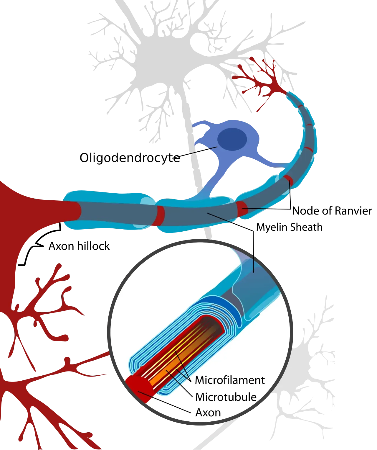
Myelin is often described as the brain’s insulation. It wraps around nerve fibers, helping electrical signals travel rapidly and efficiently. But scientists are increasingly finding that myelin is far more dynamic than a simple protective coating. A new research perspective proposes that myelin may behave like a responsive biological material, changing its structure and physical properties in response to electrical activity, hydration, mechanical forces and other signals.
Myelin is made of many tightly packed layers of lipid-rich membranes. These layers can swell, compress and reorganize. Researchers believe that changes in the water between these layers may be particularly important because hydration can influence the stiffness, electrical properties and stability of the myelin structure. The study also explores an intriguing possibility: myelin may retain a physical “memory” of previous stimulation. This does not mean myelin stores memories in the same way the brain stores experiences. Rather, researchers propose that changes in membrane structure and energy may persist after stimulation has ended, potentially affecting how the material responds to later signals.To investigate this idea, scientists are combining several advanced techniques, including electrical measurements, neutron and X-ray scattering, mechanical measurements and terahertz spectroscopy. These methods allow researchers to examine myelin-like membranes from different perspectives—from their molecular structure to their electrical and mechanical behavior.
The work is especially relevant to diseases involving myelin damage, including multiple sclerosis and other demyelinating disorders. Early myelin damage can involve swelling and changes in organization before more permanent deterioration occurs. Understanding these early physical changes could eventually help scientists identify damage sooner and explore ways to preserve or restore myelin.
Importantly, the researchers emphasize that the proposed memory-like behavior of myelin remains a hypothesis that requires experimental testing. The goal is to determine whether electrical stimulation, hydration and structural changes can produce measurable, history-dependent responses. If confirmed, the findings could change how scientists think about myelin—from a passive insulation system to an adaptive structure that participates in the brain’s complex exchange of electrical, mechanical and metabolic energy.
The research, supported by the National Science Foundation, also points beyond neuroscience. Understanding how biological membranes store and redistribute energy could inspire new materials for sensing, computing and other adaptive technologies.
In short, the research raises an important question: Could the physical behavior of myelin provide another piece of the puzzle of how the nervous system adapts—and how early damage might be detected before it becomes irreversible?
Healthy Myelin → Electrical Signal → Structural Changes → Early Myelin Damage
Abstract — Neuralink, Brain-Computer Interfaces and Parkinson’s Disease
NEW BRAIN-COMPUTER TECHNOLOGY COULD OPEN NEW POSSIBILITIES FOR PARKINSON’S TREATMENT.
Brain-computer interfaces are emerging as a potential new tool for understanding and treating neurological disorders, including Parkinson’s disease. Neuralink’s implant uses tiny electrode threads to record brain activity and translate neural signals into commands for external devices, although its current clinical trials are focused primarily on paralysis rather than Parkinson’s disease. is the text area for this paragraph. To change it, simply click and start typing. Once you've added your content, you can customize its design by using different colors, fonts, font sizes and bullets. Just highlight the words you want to design and choose from the various options in the text editing bar.
Parkinson’s research, meanwhile, is showing how abnormal electrical activity in motor circuits—particularly excessive beta-band synchronization—can interfere with movement. New experimental approaches are combining brain-computer interfaces with deep brain stimulation to detect and respond to these abnormal signals.
Together, these developments raise an important possibility: future implanted brain-computer systems could potentially read abnormal movement-related brain signals and deliver personalized stimulation in real time. Such technology remains experimental, and Neuralink itself has not yet demonstrated a Parkinson’s treatment in a registered clinical trial. Nevertheless, the convergence of BCI technology, neural recording and adaptive stimulation could open a new direction for Parkinson’s research.